IL-37 Monoclonal Antibody (37D12), PE, eBioscience
PRODUCT DETAILS
Host: Mouse
Isotype: IgG1, kappa
Clonality: Monoclonal
Clone: 37D12
Format: PE
Reactivity: Hu
Application: Flow Cytometry
Tested Dilution: 5 µL (0.06 µg)/test
Concentration: 5 μL/Test
Storage: 4°C, store in dark, DO NOT FREEZE!
Formulation: PBS with BSA and 0.09% sodium azide; pH 7.2
Purification: Affinity chromatography
Data Sheet: TDS
Specific Information
Description: This 37D12 monoclonal antibody reacts with human IL-37. IL-37 is a member of the interleukin 1 cytokine family and is also known as IL-1F7 and IL-1H4. IL-37 suppresses innate inflammatory and immune responses and exhibits anti-inflammatory characteristics resembling those of IL-1R8. IL-37 binds to the alpha chain of IL-18R with low affinity and does not recruit the essential signaling co-receptor IL-18 beta or mediate pro-inflammatory activity. IL-37 also binds to IL-18 binding protein (IL-18BP) and enhances the antagonistic effects of IL-18BP. IL-37 forms a trimeric complex with IL-18BP and IL-18R beta, which results in the blockade of IL-18 activity. Transcripts of IL-37 have been detected in human tissues, such as lung, testis, and colon tumors, as well as some human cell lines.
Applications Reported: This 37D12 antibody has been reported for use in intracellular staining followed by flow cytometric analysis.
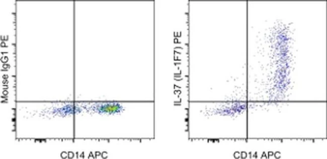
Applications Tested: This 37D12 antibody has been pre-titrated and tested by intracellular staining followed by flow cytometric analysis of normal human peripheral blood cells using the Intracellular Fixation & Permeabilization Buffer Set (Product # 88-8824-00) and protocol. Please refer to BestProtocols®: Protocol A: Two step protocol for (cytoplasmic) intracellular proteins located under the Resources Tab online. This can be used at 5 µL (0.06 µg) per test. A test is defined as the amount (µg) of antibody that will stain a cell sample in a final volume of 100 µL. Cell number should be determined empirically but can range from 10^5 to 10^8 cells/test.
Excitation: 488-561 nm; Emission: 578 nm; Laser: Blue Laser, Green Laser, Yellow-Green Laser.
Filtration: 0.2 µm post-manufacturing filtered.
For Research Use Only. Not for use in diagnostic procedures. Not for resale without express authorization.
Product Information
Product Information
Shipping & Returns
Shipping & Returns
IL-37 Monoclonal Antibody (37D12), PE, eBioscience
IL-37 Monoclonal Antibody (37D12), PE, eBioscience
PRODUCT DETAILS
Host: Mouse
Isotype: IgG1, kappa
Clonality: Monoclonal
Clone: 37D12
Format: PE
Reactivity: Hu
Application: Flow Cytometry
Tested Dilution: 5 µL (0.06 µg)/test
Concentration: 5 μL/Test
Storage: 4°C, store in dark, DO NOT FREEZE!
Formulation: PBS with BSA and 0.09% sodium azide; pH 7.2
Purification: Affinity chromatography
Data Sheet: TDS
Specific Information
Description: This 37D12 monoclonal antibody reacts with human IL-37. IL-37 is a member of the interleukin 1 cytokine family and is also known as IL-1F7 and IL-1H4. IL-37 suppresses innate inflammatory and immune responses and exhibits anti-inflammatory characteristics resembling those of IL-1R8. IL-37 binds to the alpha chain of IL-18R with low affinity and does not recruit the essential signaling co-receptor IL-18 beta or mediate pro-inflammatory activity. IL-37 also binds to IL-18 binding protein (IL-18BP) and enhances the antagonistic effects of IL-18BP. IL-37 forms a trimeric complex with IL-18BP and IL-18R beta, which results in the blockade of IL-18 activity. Transcripts of IL-37 have been detected in human tissues, such as lung, testis, and colon tumors, as well as some human cell lines.
Applications Reported: This 37D12 antibody has been reported for use in intracellular staining followed by flow cytometric analysis.
Applications Tested: This 37D12 antibody has been pre-titrated and tested by intracellular staining followed by flow cytometric analysis of normal human peripheral blood cells using the Intracellular Fixation & Permeabilization Buffer Set (Product # 88-8824-00) and protocol. Please refer to BestProtocols®: Protocol A: Two step protocol for (cytoplasmic) intracellular proteins located under the Resources Tab online. This can be used at 5 µL (0.06 µg) per test. A test is defined as the amount (µg) of antibody that will stain a cell sample in a final volume of 100 µL. Cell number should be determined empirically but can range from 10^5 to 10^8 cells/test.
Excitation: 488-561 nm; Emission: 578 nm; Laser: Blue Laser, Green Laser, Yellow-Green Laser.
Filtration: 0.2 µm post-manufacturing filtered.
For Research Use Only. Not for use in diagnostic procedures. Not for resale without express authorization.
Product Information
Product Information
Shipping & Returns
Shipping & Returns
Description
PRODUCT DETAILS
Host: Mouse
Isotype: IgG1, kappa
Clonality: Monoclonal
Clone: 37D12
Format: PE
Reactivity: Hu
Application: Flow Cytometry
Tested Dilution: 5 µL (0.06 µg)/test
Concentration: 5 μL/Test
Storage: 4°C, store in dark, DO NOT FREEZE!
Formulation: PBS with BSA and 0.09% sodium azide; pH 7.2
Purification: Affinity chromatography
Data Sheet: TDS
Specific Information
Description: This 37D12 monoclonal antibody reacts with human IL-37. IL-37 is a member of the interleukin 1 cytokine family and is also known as IL-1F7 and IL-1H4. IL-37 suppresses innate inflammatory and immune responses and exhibits anti-inflammatory characteristics resembling those of IL-1R8. IL-37 binds to the alpha chain of IL-18R with low affinity and does not recruit the essential signaling co-receptor IL-18 beta or mediate pro-inflammatory activity. IL-37 also binds to IL-18 binding protein (IL-18BP) and enhances the antagonistic effects of IL-18BP. IL-37 forms a trimeric complex with IL-18BP and IL-18R beta, which results in the blockade of IL-18 activity. Transcripts of IL-37 have been detected in human tissues, such as lung, testis, and colon tumors, as well as some human cell lines.
Applications Reported: This 37D12 antibody has been reported for use in intracellular staining followed by flow cytometric analysis.
Applications Tested: This 37D12 antibody has been pre-titrated and tested by intracellular staining followed by flow cytometric analysis of normal human peripheral blood cells using the Intracellular Fixation & Permeabilization Buffer Set (Product # 88-8824-00) and protocol. Please refer to BestProtocols®: Protocol A: Two step protocol for (cytoplasmic) intracellular proteins located under the Resources Tab online. This can be used at 5 µL (0.06 µg) per test. A test is defined as the amount (µg) of antibody that will stain a cell sample in a final volume of 100 µL. Cell number should be determined empirically but can range from 10^5 to 10^8 cells/test.
Excitation: 488-561 nm; Emission: 578 nm; Laser: Blue Laser, Green Laser, Yellow-Green Laser.
Filtration: 0.2 µm post-manufacturing filtered.
For Research Use Only. Not for use in diagnostic procedures. Not for resale without express authorization.


