IL-23 p19 Monoclonal Antibody (23dcdp), PE, eBioscience
PRODUCT DETAILS
Host: Mouse
Isotype: IgG2b, kappa
Clonality: Monoclonal
Clone: 23dcdp
Format: PE
Reactivity: Hu
Application: Flow Cytometry
Tested Dilution: 5 µL (0.06 µg)/test
Concentration: 5 μL/Test
Storage: 4°C, store in dark, DO NOT FREEZE!
Formulation: PBS with BSA and 0.09% sodium azide; pH 7.2
Purification: Affinity chromatography
Data Sheet: TDS
Specific Information
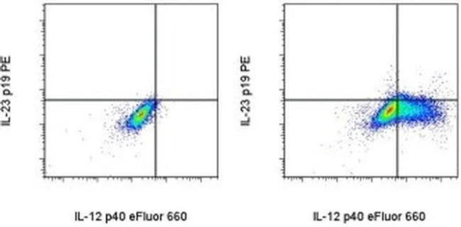
Description: This 23dcdp monoclonal antibody reacts with the p19 subunit of human IL-23. This heterodimeric cytokine is composed of two disulfide-linked subunits, p40 and p19. It is closely related to IL-12, with which it shares the p40 subunit. The IL-23 receptor is also heterodimeric and shares the IL-12Rbeta1 chain with IL-12, while the IL-23R chain is unique to the IL-23 receptor complex. IL-23R signaling occurs through the Jak/STAT pathway and results in RORgammat expression, which promotes maintenance and proliferation of T helper 17 (Th17) cells.
Dendritic cells and macrophages produce IL-23 in response to TLR2, TLR4, and TLR8 ligands, as well as agonists of the beta-glucan receptor, Dectin-1. Our studies suggest donor, kinetics and type of stimulant can result in variation in IL-23 expression levels. Recent publications suggest the p19 subunit may also exist in the absence of association with p40.
Applications Reported: This 23dcdp antibody has been reported for use in intracellular staining followed by flow cytometric analysis.
Applications Tested: This 23dcdp antibody has been pre-titrated and tested by intracellular staining followed by flow cytometric analysis. This can be used at 5 µL (0.06 µg) per test. A test is defined as the amount (µg) of antibody that will stain a cell sample in a final volume of 100 µL. Cell number should be determined empirically but can range from 10^5 to 10^8 cells/test.
Excitation: 488-561 nm; Emission: 578 nm; Laser: Blue Laser, Green Laser, Yellow-Green Laser.
Filtration: 0.2 µm post-manufacturing filtered.
For Research Use Only. Not for use in diagnostic procedures. Not for resale without express authorization.
Product Information
Product Information
Shipping & Returns
Shipping & Returns
IL-23 p19 Monoclonal Antibody (23dcdp), PE, eBioscience
IL-23 p19 Monoclonal Antibody (23dcdp), PE, eBioscience
PRODUCT DETAILS
Host: Mouse
Isotype: IgG2b, kappa
Clonality: Monoclonal
Clone: 23dcdp
Format: PE
Reactivity: Hu
Application: Flow Cytometry
Tested Dilution: 5 µL (0.06 µg)/test
Concentration: 5 μL/Test
Storage: 4°C, store in dark, DO NOT FREEZE!
Formulation: PBS with BSA and 0.09% sodium azide; pH 7.2
Purification: Affinity chromatography
Data Sheet: TDS
Specific Information
Description: This 23dcdp monoclonal antibody reacts with the p19 subunit of human IL-23. This heterodimeric cytokine is composed of two disulfide-linked subunits, p40 and p19. It is closely related to IL-12, with which it shares the p40 subunit. The IL-23 receptor is also heterodimeric and shares the IL-12Rbeta1 chain with IL-12, while the IL-23R chain is unique to the IL-23 receptor complex. IL-23R signaling occurs through the Jak/STAT pathway and results in RORgammat expression, which promotes maintenance and proliferation of T helper 17 (Th17) cells.
Dendritic cells and macrophages produce IL-23 in response to TLR2, TLR4, and TLR8 ligands, as well as agonists of the beta-glucan receptor, Dectin-1. Our studies suggest donor, kinetics and type of stimulant can result in variation in IL-23 expression levels. Recent publications suggest the p19 subunit may also exist in the absence of association with p40.
Applications Reported: This 23dcdp antibody has been reported for use in intracellular staining followed by flow cytometric analysis.
Applications Tested: This 23dcdp antibody has been pre-titrated and tested by intracellular staining followed by flow cytometric analysis. This can be used at 5 µL (0.06 µg) per test. A test is defined as the amount (µg) of antibody that will stain a cell sample in a final volume of 100 µL. Cell number should be determined empirically but can range from 10^5 to 10^8 cells/test.
Excitation: 488-561 nm; Emission: 578 nm; Laser: Blue Laser, Green Laser, Yellow-Green Laser.
Filtration: 0.2 µm post-manufacturing filtered.
For Research Use Only. Not for use in diagnostic procedures. Not for resale without express authorization.
Original: $460.00
-70%$460.00
$138.00Product Information
Product Information
Shipping & Returns
Shipping & Returns
Description
PRODUCT DETAILS
Host: Mouse
Isotype: IgG2b, kappa
Clonality: Monoclonal
Clone: 23dcdp
Format: PE
Reactivity: Hu
Application: Flow Cytometry
Tested Dilution: 5 µL (0.06 µg)/test
Concentration: 5 μL/Test
Storage: 4°C, store in dark, DO NOT FREEZE!
Formulation: PBS with BSA and 0.09% sodium azide; pH 7.2
Purification: Affinity chromatography
Data Sheet: TDS
Specific Information
Description: This 23dcdp monoclonal antibody reacts with the p19 subunit of human IL-23. This heterodimeric cytokine is composed of two disulfide-linked subunits, p40 and p19. It is closely related to IL-12, with which it shares the p40 subunit. The IL-23 receptor is also heterodimeric and shares the IL-12Rbeta1 chain with IL-12, while the IL-23R chain is unique to the IL-23 receptor complex. IL-23R signaling occurs through the Jak/STAT pathway and results in RORgammat expression, which promotes maintenance and proliferation of T helper 17 (Th17) cells.
Dendritic cells and macrophages produce IL-23 in response to TLR2, TLR4, and TLR8 ligands, as well as agonists of the beta-glucan receptor, Dectin-1. Our studies suggest donor, kinetics and type of stimulant can result in variation in IL-23 expression levels. Recent publications suggest the p19 subunit may also exist in the absence of association with p40.
Applications Reported: This 23dcdp antibody has been reported for use in intracellular staining followed by flow cytometric analysis.
Applications Tested: This 23dcdp antibody has been pre-titrated and tested by intracellular staining followed by flow cytometric analysis. This can be used at 5 µL (0.06 µg) per test. A test is defined as the amount (µg) of antibody that will stain a cell sample in a final volume of 100 µL. Cell number should be determined empirically but can range from 10^5 to 10^8 cells/test.
Excitation: 488-561 nm; Emission: 578 nm; Laser: Blue Laser, Green Laser, Yellow-Green Laser.
Filtration: 0.2 µm post-manufacturing filtered.
For Research Use Only. Not for use in diagnostic procedures. Not for resale without express authorization.


