IDO Monoclonal Antibody (eyedio), APC-eFluor 780, eBioscience
PRODUCT DETAILS
Host: Mouse
Isotype: IgG1, kappa
Clonality: Monoclonal
Clone: eyedio
Format: APC-eFluor 780
Reactivity: Hu
Application: Flow Cytometry
Tested Dilution: 5 µL (0.125 µg)/test
Concentration: 5 μL/Test
Storage: 4°C, store in dark, DO NOT FREEZE!
Formulation: PBS with BSA and 0.09% sodium azide; pH 7.2
Purification: Affinity chromatography
Data Sheet: TDS
Specific Information
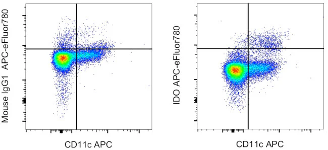
Description: This eyedio monoclonal antibody reacts with human indoleamine-2,3-dioxygenase (IDO, INDO, IDO1), an intracellular enzyme that catalyzes the degradation of tryptophan to kynurenines. IDO is expressed in a wide variety of tissues and cells, including macrophages, plasmacytoid dendritic cells, and several cell lines. IDO can be induced in many different cell types by IFN gamma or other inflammatory stimuli. Expression of IDO in antigen presenting cells and tumors is thought to mediate immune suppression through depletion of this essential amino acid and/or by the creation of tryptophan metabolites that cause apoptosis of T cells and induction of regulatory T cells.
Applications Reported: This eyedio antibody has been reported for use in flow cytometric analysis.
Applications Tested: This eyedio antibody has been pre-diluted and tested by flow cytometric analysis of normal human peripheral blood cells using the Intracellular Fixation & Permeabilization Buffer Set (Product # 88-8824-00) and protocol. Please refer to "Staining Intracellular Antigens for Flow Cytometry, Protocol A: Two step protocol for intracellular (cytoplasmic) proteins" located at thermofisher.com. This may be used at 5 µL (0.125 µg) per test. A test is defined as the amount (µg) of antibody that will stain a cell sample in a final volume of 100 µL. Cell number should be determined empirically but can range from 10^5 to 10^8 cells/test.
APC-eFluor 780 emits at 780 nm and is excited with the Red laser (633-647 nm). Please make sure that your instrument is capable of detecting this fluorochrome.
Light sensitivity: This tandem dye is sensitive to photo-induced oxidation. Please protect this vial and stained samples from light.
Fixation: Samples can be stored in IC Fixation Buffer (Product # 00-8222-49) (100 µL of cell sample + 100 µL of IC Fixation Buffer) or 1-step Fix/Lyse Solution (Product # 00-5333-57) for up to 3 days in the dark at 4°C with minimal impact on brightness and FRET efficiency/compensation. Some generalizations regarding fluorophore performance after fixation can be made, but clone specific performance should be determined empirically.
Excitation: 633-647 nm; Emission: 780 nm; Laser: Red Laser.
For Research Use Only. Not for use in diagnostic procedures. Not for resale without express authorization.
Product Information
Product Information
Shipping & Returns
Shipping & Returns
IDO Monoclonal Antibody (eyedio), APC-eFluor 780, eBioscience
IDO Monoclonal Antibody (eyedio), APC-eFluor 780, eBioscience
PRODUCT DETAILS
Host: Mouse
Isotype: IgG1, kappa
Clonality: Monoclonal
Clone: eyedio
Format: APC-eFluor 780
Reactivity: Hu
Application: Flow Cytometry
Tested Dilution: 5 µL (0.125 µg)/test
Concentration: 5 μL/Test
Storage: 4°C, store in dark, DO NOT FREEZE!
Formulation: PBS with BSA and 0.09% sodium azide; pH 7.2
Purification: Affinity chromatography
Data Sheet: TDS
Specific Information
Description: This eyedio monoclonal antibody reacts with human indoleamine-2,3-dioxygenase (IDO, INDO, IDO1), an intracellular enzyme that catalyzes the degradation of tryptophan to kynurenines. IDO is expressed in a wide variety of tissues and cells, including macrophages, plasmacytoid dendritic cells, and several cell lines. IDO can be induced in many different cell types by IFN gamma or other inflammatory stimuli. Expression of IDO in antigen presenting cells and tumors is thought to mediate immune suppression through depletion of this essential amino acid and/or by the creation of tryptophan metabolites that cause apoptosis of T cells and induction of regulatory T cells.
Applications Reported: This eyedio antibody has been reported for use in flow cytometric analysis.
Applications Tested: This eyedio antibody has been pre-diluted and tested by flow cytometric analysis of normal human peripheral blood cells using the Intracellular Fixation & Permeabilization Buffer Set (Product # 88-8824-00) and protocol. Please refer to "Staining Intracellular Antigens for Flow Cytometry, Protocol A: Two step protocol for intracellular (cytoplasmic) proteins" located at thermofisher.com. This may be used at 5 µL (0.125 µg) per test. A test is defined as the amount (µg) of antibody that will stain a cell sample in a final volume of 100 µL. Cell number should be determined empirically but can range from 10^5 to 10^8 cells/test.
APC-eFluor 780 emits at 780 nm and is excited with the Red laser (633-647 nm). Please make sure that your instrument is capable of detecting this fluorochrome.
Light sensitivity: This tandem dye is sensitive to photo-induced oxidation. Please protect this vial and stained samples from light.
Fixation: Samples can be stored in IC Fixation Buffer (Product # 00-8222-49) (100 µL of cell sample + 100 µL of IC Fixation Buffer) or 1-step Fix/Lyse Solution (Product # 00-5333-57) for up to 3 days in the dark at 4°C with minimal impact on brightness and FRET efficiency/compensation. Some generalizations regarding fluorophore performance after fixation can be made, but clone specific performance should be determined empirically.
Excitation: 633-647 nm; Emission: 780 nm; Laser: Red Laser.
For Research Use Only. Not for use in diagnostic procedures. Not for resale without express authorization.
Original: $485.00
-70%$485.00
$145.50Product Information
Product Information
Shipping & Returns
Shipping & Returns
Description
PRODUCT DETAILS
Host: Mouse
Isotype: IgG1, kappa
Clonality: Monoclonal
Clone: eyedio
Format: APC-eFluor 780
Reactivity: Hu
Application: Flow Cytometry
Tested Dilution: 5 µL (0.125 µg)/test
Concentration: 5 μL/Test
Storage: 4°C, store in dark, DO NOT FREEZE!
Formulation: PBS with BSA and 0.09% sodium azide; pH 7.2
Purification: Affinity chromatography
Data Sheet: TDS
Specific Information
Description: This eyedio monoclonal antibody reacts with human indoleamine-2,3-dioxygenase (IDO, INDO, IDO1), an intracellular enzyme that catalyzes the degradation of tryptophan to kynurenines. IDO is expressed in a wide variety of tissues and cells, including macrophages, plasmacytoid dendritic cells, and several cell lines. IDO can be induced in many different cell types by IFN gamma or other inflammatory stimuli. Expression of IDO in antigen presenting cells and tumors is thought to mediate immune suppression through depletion of this essential amino acid and/or by the creation of tryptophan metabolites that cause apoptosis of T cells and induction of regulatory T cells.
Applications Reported: This eyedio antibody has been reported for use in flow cytometric analysis.
Applications Tested: This eyedio antibody has been pre-diluted and tested by flow cytometric analysis of normal human peripheral blood cells using the Intracellular Fixation & Permeabilization Buffer Set (Product # 88-8824-00) and protocol. Please refer to "Staining Intracellular Antigens for Flow Cytometry, Protocol A: Two step protocol for intracellular (cytoplasmic) proteins" located at thermofisher.com. This may be used at 5 µL (0.125 µg) per test. A test is defined as the amount (µg) of antibody that will stain a cell sample in a final volume of 100 µL. Cell number should be determined empirically but can range from 10^5 to 10^8 cells/test.
APC-eFluor 780 emits at 780 nm and is excited with the Red laser (633-647 nm). Please make sure that your instrument is capable of detecting this fluorochrome.
Light sensitivity: This tandem dye is sensitive to photo-induced oxidation. Please protect this vial and stained samples from light.
Fixation: Samples can be stored in IC Fixation Buffer (Product # 00-8222-49) (100 µL of cell sample + 100 µL of IC Fixation Buffer) or 1-step Fix/Lyse Solution (Product # 00-5333-57) for up to 3 days in the dark at 4°C with minimal impact on brightness and FRET efficiency/compensation. Some generalizations regarding fluorophore performance after fixation can be made, but clone specific performance should be determined empirically.
Excitation: 633-647 nm; Emission: 780 nm; Laser: Red Laser.
For Research Use Only. Not for use in diagnostic procedures. Not for resale without express authorization.


