IDO Monoclonal Antibody (eyedio), Alexa Fluor 700, eBioscience
PRODUCT DETAILS
Host: Mouse
Isotype: IgG1, kappa
Clonality: Monoclonal
Clone: eyedio
Format: Alexa Fluor 700
Reactivity: Hu
Application: Flow Cytometry
Tested Dilution: 5 µL (0.125 µg)/test
Concentration: 5 μL/Test
Storage: 4°C, store in dark, DO NOT FREEZE!
Formulation: PBS with BSA and 0.09% sodium azide; pH 7.2
Purification: Affinity chromatography
Data Sheet: TDS
Specific Information
Description: This eyedio monoclonal antibody reacts with human indoleamine-2, 3-dioxygenase (IDO, INDO, IDO1), an intracellular enzyme that catalyzes the degradation of tryptophan to kynurenines. IDO is expressed in a wide variety of tissues and cells, including macrophages, plasmacytoid dendritic cells, and several cell lines. IDO can be induced in many different cell types by IFN gamma or other inflammatory stimuli. Expression of IDO in antigen presenting cells and tumors is thought to mediate immune suppression through depletion of this essential amino acid and/or by the creation of tryptophan metabolites that cause apoptosis of T cells and induction of regulatory T cells.
Applications Reported: This eyedio antibody has been reported for use in intracellular staining followed by flow cytometric analysis.
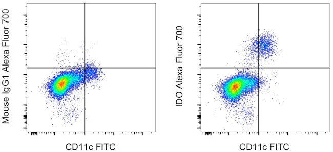
Applications Tested: This eyedio antibody has been pre-diluted and tested by intracellular staining followed flow cytometric analysis of stimulated normal human peripheral blood cells using the Intracellular Fixation & Permeabilization Buffer Set (Product # 88-8824-00) and protocol. Please refer to "Staining Intracellular Antigens for Flow Cytometry, Protocol A: Two step protocol for intracellular (cytoplasmic) proteins" located at www.thermofisher.com/flowprotocols . This may be used at 5 µL (0.125 µg) per test. A test is defined as the amount (µg) of antibody that will stain a cell sample in a final volume of 100 µL. Cell number should be determined empirically but can range from 10^5 to 10^8 cells/test.
Alexa Fluor 700 emits at 723 nm and can be excited with the red laser (633-647 nm). Most instruments will require a 685 LP mirror and 710/20 band pass filter. Please make sure that your instrument is capable of detecting this fluorochrome.
Excitation: 633-647 nm; Emission: 723 nm; Laser: Red Laser.
For Research Use Only. Not for use in diagnostic procedures. Not for resale without express authorization.
Product Information
Product Information
Shipping & Returns
Shipping & Returns
IDO Monoclonal Antibody (eyedio), Alexa Fluor 700, eBioscience
IDO Monoclonal Antibody (eyedio), Alexa Fluor 700, eBioscience
PRODUCT DETAILS
Host: Mouse
Isotype: IgG1, kappa
Clonality: Monoclonal
Clone: eyedio
Format: Alexa Fluor 700
Reactivity: Hu
Application: Flow Cytometry
Tested Dilution: 5 µL (0.125 µg)/test
Concentration: 5 μL/Test
Storage: 4°C, store in dark, DO NOT FREEZE!
Formulation: PBS with BSA and 0.09% sodium azide; pH 7.2
Purification: Affinity chromatography
Data Sheet: TDS
Specific Information
Description: This eyedio monoclonal antibody reacts with human indoleamine-2, 3-dioxygenase (IDO, INDO, IDO1), an intracellular enzyme that catalyzes the degradation of tryptophan to kynurenines. IDO is expressed in a wide variety of tissues and cells, including macrophages, plasmacytoid dendritic cells, and several cell lines. IDO can be induced in many different cell types by IFN gamma or other inflammatory stimuli. Expression of IDO in antigen presenting cells and tumors is thought to mediate immune suppression through depletion of this essential amino acid and/or by the creation of tryptophan metabolites that cause apoptosis of T cells and induction of regulatory T cells.
Applications Reported: This eyedio antibody has been reported for use in intracellular staining followed by flow cytometric analysis.
Applications Tested: This eyedio antibody has been pre-diluted and tested by intracellular staining followed flow cytometric analysis of stimulated normal human peripheral blood cells using the Intracellular Fixation & Permeabilization Buffer Set (Product # 88-8824-00) and protocol. Please refer to "Staining Intracellular Antigens for Flow Cytometry, Protocol A: Two step protocol for intracellular (cytoplasmic) proteins" located at www.thermofisher.com/flowprotocols . This may be used at 5 µL (0.125 µg) per test. A test is defined as the amount (µg) of antibody that will stain a cell sample in a final volume of 100 µL. Cell number should be determined empirically but can range from 10^5 to 10^8 cells/test.
Alexa Fluor 700 emits at 723 nm and can be excited with the red laser (633-647 nm). Most instruments will require a 685 LP mirror and 710/20 band pass filter. Please make sure that your instrument is capable of detecting this fluorochrome.
Excitation: 633-647 nm; Emission: 723 nm; Laser: Red Laser.
For Research Use Only. Not for use in diagnostic procedures. Not for resale without express authorization.
Original: $423.00
-70%$423.00
$126.90Product Information
Product Information
Shipping & Returns
Shipping & Returns
Description
PRODUCT DETAILS
Host: Mouse
Isotype: IgG1, kappa
Clonality: Monoclonal
Clone: eyedio
Format: Alexa Fluor 700
Reactivity: Hu
Application: Flow Cytometry
Tested Dilution: 5 µL (0.125 µg)/test
Concentration: 5 μL/Test
Storage: 4°C, store in dark, DO NOT FREEZE!
Formulation: PBS with BSA and 0.09% sodium azide; pH 7.2
Purification: Affinity chromatography
Data Sheet: TDS
Specific Information
Description: This eyedio monoclonal antibody reacts with human indoleamine-2, 3-dioxygenase (IDO, INDO, IDO1), an intracellular enzyme that catalyzes the degradation of tryptophan to kynurenines. IDO is expressed in a wide variety of tissues and cells, including macrophages, plasmacytoid dendritic cells, and several cell lines. IDO can be induced in many different cell types by IFN gamma or other inflammatory stimuli. Expression of IDO in antigen presenting cells and tumors is thought to mediate immune suppression through depletion of this essential amino acid and/or by the creation of tryptophan metabolites that cause apoptosis of T cells and induction of regulatory T cells.
Applications Reported: This eyedio antibody has been reported for use in intracellular staining followed by flow cytometric analysis.
Applications Tested: This eyedio antibody has been pre-diluted and tested by intracellular staining followed flow cytometric analysis of stimulated normal human peripheral blood cells using the Intracellular Fixation & Permeabilization Buffer Set (Product # 88-8824-00) and protocol. Please refer to "Staining Intracellular Antigens for Flow Cytometry, Protocol A: Two step protocol for intracellular (cytoplasmic) proteins" located at www.thermofisher.com/flowprotocols . This may be used at 5 µL (0.125 µg) per test. A test is defined as the amount (µg) of antibody that will stain a cell sample in a final volume of 100 µL. Cell number should be determined empirically but can range from 10^5 to 10^8 cells/test.
Alexa Fluor 700 emits at 723 nm and can be excited with the red laser (633-647 nm). Most instruments will require a 685 LP mirror and 710/20 band pass filter. Please make sure that your instrument is capable of detecting this fluorochrome.
Excitation: 633-647 nm; Emission: 723 nm; Laser: Red Laser.
For Research Use Only. Not for use in diagnostic procedures. Not for resale without express authorization.


