CD49b (Integrin alpha 2) Monoclonal Antibody (P1H5), PE, eBioscience
PRODUCT DETAILS
Host: Mouse
Isotype: IgG2b, kappa
Clonality: Monoclonal
Clone: P1H5
Format: PE
Reactivity: Hu
Application: Flow Cytometry
Tested Dilution: 5 µL (0.25 µg)/test
Concentration: 5 μL/Test
Storage: 4°C, store in dark, DO NOT FREEZE!
Formulation: PBS with BSA and 0.09% sodium azide; pH 7.2
Purification: Affinity chromatography
Data Sheet: TDS
Specific Information
Description: This P1H5 monoclonal antibody reacts with human CD49b, which is also known as integrin alpha 2. Integrins constitute a family of cell surface receptors that mediate adhesion between cells or between a cell and the extracellular matrix. These proteins are heterodimers consisting of two distinct subunits, alpha and beta. Integrins are expressed on hematopoietic, non-hematopoietic, and cancer cells. Integrin-mediated adhesion is regulated by changes in ligand affinity (or activation), cell shape, and integrin clustering and diffusion. Integrin alpha 2 forms a complex with the common beta 1 subunit to form the alpha2beta1 integrin, which binds type I collagen and laminin. Moreover, studies suggest that this complex suppresses metastasis in cancer and participates in T cell costimulation by activating the MAPK and PI3K pathways.
The P1H5 monoclonal antibody has been reported to inhibit adhesion of fibroblasts and platelets to collagen. Crossblocking studies indicate that P1H5 and eBioY418 recognize different epitopes.
Applications Reported: This P1H5 antibody has been reported for use in flow cytometric analysis.
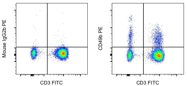
Applications Tested: This P1H5 antibody has been pre-diluted and tested by flow cytometric analysis of normal human peripheral blood cells. This may be used at 5 µL (0.25 µg) per test. A test is defined as the amount (µg) of antibody that will stain a cell sample in a final volume of 100 µL. Cell number should be determined empirically but can range from 10^5 to 10^8 cells/test.
Excitation: 488-561 nm; Emission: 578 nm; Laser: Blue Laser, Green Laser, Yellow-Green Laser
For Research Use Only. Not for use in diagnostic procedures. Not for resale without express authorization.
Product Information
Product Information
Shipping & Returns
Shipping & Returns
CD49b (Integrin alpha 2) Monoclonal Antibody (P1H5), PE, eBioscience
CD49b (Integrin alpha 2) Monoclonal Antibody (P1H5), PE, eBioscience
PRODUCT DETAILS
Host: Mouse
Isotype: IgG2b, kappa
Clonality: Monoclonal
Clone: P1H5
Format: PE
Reactivity: Hu
Application: Flow Cytometry
Tested Dilution: 5 µL (0.25 µg)/test
Concentration: 5 μL/Test
Storage: 4°C, store in dark, DO NOT FREEZE!
Formulation: PBS with BSA and 0.09% sodium azide; pH 7.2
Purification: Affinity chromatography
Data Sheet: TDS
Specific Information
Description: This P1H5 monoclonal antibody reacts with human CD49b, which is also known as integrin alpha 2. Integrins constitute a family of cell surface receptors that mediate adhesion between cells or between a cell and the extracellular matrix. These proteins are heterodimers consisting of two distinct subunits, alpha and beta. Integrins are expressed on hematopoietic, non-hematopoietic, and cancer cells. Integrin-mediated adhesion is regulated by changes in ligand affinity (or activation), cell shape, and integrin clustering and diffusion. Integrin alpha 2 forms a complex with the common beta 1 subunit to form the alpha2beta1 integrin, which binds type I collagen and laminin. Moreover, studies suggest that this complex suppresses metastasis in cancer and participates in T cell costimulation by activating the MAPK and PI3K pathways.
The P1H5 monoclonal antibody has been reported to inhibit adhesion of fibroblasts and platelets to collagen. Crossblocking studies indicate that P1H5 and eBioY418 recognize different epitopes.
Applications Reported: This P1H5 antibody has been reported for use in flow cytometric analysis.
Applications Tested: This P1H5 antibody has been pre-diluted and tested by flow cytometric analysis of normal human peripheral blood cells. This may be used at 5 µL (0.25 µg) per test. A test is defined as the amount (µg) of antibody that will stain a cell sample in a final volume of 100 µL. Cell number should be determined empirically but can range from 10^5 to 10^8 cells/test.
Excitation: 488-561 nm; Emission: 578 nm; Laser: Blue Laser, Green Laser, Yellow-Green Laser
For Research Use Only. Not for use in diagnostic procedures. Not for resale without express authorization.
Original: $370.00
-70%$370.00
$111.00Product Information
Product Information
Shipping & Returns
Shipping & Returns
Description
PRODUCT DETAILS
Host: Mouse
Isotype: IgG2b, kappa
Clonality: Monoclonal
Clone: P1H5
Format: PE
Reactivity: Hu
Application: Flow Cytometry
Tested Dilution: 5 µL (0.25 µg)/test
Concentration: 5 μL/Test
Storage: 4°C, store in dark, DO NOT FREEZE!
Formulation: PBS with BSA and 0.09% sodium azide; pH 7.2
Purification: Affinity chromatography
Data Sheet: TDS
Specific Information
Description: This P1H5 monoclonal antibody reacts with human CD49b, which is also known as integrin alpha 2. Integrins constitute a family of cell surface receptors that mediate adhesion between cells or between a cell and the extracellular matrix. These proteins are heterodimers consisting of two distinct subunits, alpha and beta. Integrins are expressed on hematopoietic, non-hematopoietic, and cancer cells. Integrin-mediated adhesion is regulated by changes in ligand affinity (or activation), cell shape, and integrin clustering and diffusion. Integrin alpha 2 forms a complex with the common beta 1 subunit to form the alpha2beta1 integrin, which binds type I collagen and laminin. Moreover, studies suggest that this complex suppresses metastasis in cancer and participates in T cell costimulation by activating the MAPK and PI3K pathways.
The P1H5 monoclonal antibody has been reported to inhibit adhesion of fibroblasts and platelets to collagen. Crossblocking studies indicate that P1H5 and eBioY418 recognize different epitopes.
Applications Reported: This P1H5 antibody has been reported for use in flow cytometric analysis.
Applications Tested: This P1H5 antibody has been pre-diluted and tested by flow cytometric analysis of normal human peripheral blood cells. This may be used at 5 µL (0.25 µg) per test. A test is defined as the amount (µg) of antibody that will stain a cell sample in a final volume of 100 µL. Cell number should be determined empirically but can range from 10^5 to 10^8 cells/test.
Excitation: 488-561 nm; Emission: 578 nm; Laser: Blue Laser, Green Laser, Yellow-Green Laser
For Research Use Only. Not for use in diagnostic procedures. Not for resale without express authorization.


