CD39 Monoclonal Antibody (eBioA1 (A1)), FITC, eBioscience
PRODUCT DETAILS
Host: Mouse
Isotype: IgG1, kappa
Clonality: Monoclonal
Clone: eBioA1 (A1)
Format: FITC
Reactivity: Hu
Application: Flow Cytometry
Tested Dilution: 5 µL (0.25 µg)/test
Concentration: 5 μL/Test
Storage: 4°C, store in dark, DO NOT FREEZE!
Formulation: PBS with BSA and 0.09% sodium azide; pH 7.2
Purification: Affinity chromatography
Data Sheet: TDS
Specific Information
Description: The eBioA1 monoclonal antibody reacts with human CD39 also known as ectonucleoside triphosphate diphosphohydrolase 1 (ENTPD1) or NTPDase. CD39 is an integral membrane protein with two transmembrane domains and exists as a homotetramer. It is the most prominent ectoenzyme of the immune system. The function of CD39 is to effectively remove toxic extracellular ATP by converting it to ADP or AMP. CD39 is thought to work together with CD73 to hydrolyze ATP and has been well characterized on Langerhans cells. Expression of CD39 was originally identified on activated lymphocytes. Expression is also found on a subset of T cells, B cells and dendritic cells as well as weak staining on monocytes and granulocytes.
Recently, CD39 and CD73 have been found on regulatory T cells (Treg). Expression of CD39 on Treg may facilitate their entry into inflamed areas where high levels of ATP are present. Expression of CD39 on Foxp3+CD4+ cells ranges from 25-45%.
Applications Reported: This eBioA1 (A1) antibody has been reported for use in flow cytometric analysis.
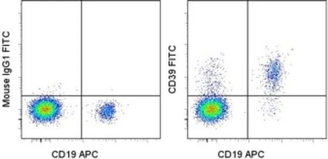
Applications Tested: This eBioA1 (A1) antibody has been pre-titrated and tested by flow cytometric analysis of normal human peripheral blood cells. This can be used at 5 µL (0.25 µg) per test. A test is defined as the amount (µg) of antibody that will stain a cell sample in a final volume of 100 µL. Cell number should be determined empirically but can range from 10^5 to 10^8 cells/test.
Excitation: 488 nm; Emission: 520 nm; Laser: Blue Laser.
Filtration: 0.2 µm post-manufacturing filtered.
For Research Use Only. Not for use in diagnostic procedures. Not for resale without express authorization.
Product Information
Product Information
Shipping & Returns
Shipping & Returns
CD39 Monoclonal Antibody (eBioA1 (A1)), FITC, eBioscience
CD39 Monoclonal Antibody (eBioA1 (A1)), FITC, eBioscience
PRODUCT DETAILS
Host: Mouse
Isotype: IgG1, kappa
Clonality: Monoclonal
Clone: eBioA1 (A1)
Format: FITC
Reactivity: Hu
Application: Flow Cytometry
Tested Dilution: 5 µL (0.25 µg)/test
Concentration: 5 μL/Test
Storage: 4°C, store in dark, DO NOT FREEZE!
Formulation: PBS with BSA and 0.09% sodium azide; pH 7.2
Purification: Affinity chromatography
Data Sheet: TDS
Specific Information
Description: The eBioA1 monoclonal antibody reacts with human CD39 also known as ectonucleoside triphosphate diphosphohydrolase 1 (ENTPD1) or NTPDase. CD39 is an integral membrane protein with two transmembrane domains and exists as a homotetramer. It is the most prominent ectoenzyme of the immune system. The function of CD39 is to effectively remove toxic extracellular ATP by converting it to ADP or AMP. CD39 is thought to work together with CD73 to hydrolyze ATP and has been well characterized on Langerhans cells. Expression of CD39 was originally identified on activated lymphocytes. Expression is also found on a subset of T cells, B cells and dendritic cells as well as weak staining on monocytes and granulocytes.
Recently, CD39 and CD73 have been found on regulatory T cells (Treg). Expression of CD39 on Treg may facilitate their entry into inflamed areas where high levels of ATP are present. Expression of CD39 on Foxp3+CD4+ cells ranges from 25-45%.
Applications Reported: This eBioA1 (A1) antibody has been reported for use in flow cytometric analysis.
Applications Tested: This eBioA1 (A1) antibody has been pre-titrated and tested by flow cytometric analysis of normal human peripheral blood cells. This can be used at 5 µL (0.25 µg) per test. A test is defined as the amount (µg) of antibody that will stain a cell sample in a final volume of 100 µL. Cell number should be determined empirically but can range from 10^5 to 10^8 cells/test.
Excitation: 488 nm; Emission: 520 nm; Laser: Blue Laser.
Filtration: 0.2 µm post-manufacturing filtered.
For Research Use Only. Not for use in diagnostic procedures. Not for resale without express authorization.
Original: $303.00
-70%$303.00
$90.90Product Information
Product Information
Shipping & Returns
Shipping & Returns
Description
PRODUCT DETAILS
Host: Mouse
Isotype: IgG1, kappa
Clonality: Monoclonal
Clone: eBioA1 (A1)
Format: FITC
Reactivity: Hu
Application: Flow Cytometry
Tested Dilution: 5 µL (0.25 µg)/test
Concentration: 5 μL/Test
Storage: 4°C, store in dark, DO NOT FREEZE!
Formulation: PBS with BSA and 0.09% sodium azide; pH 7.2
Purification: Affinity chromatography
Data Sheet: TDS
Specific Information
Description: The eBioA1 monoclonal antibody reacts with human CD39 also known as ectonucleoside triphosphate diphosphohydrolase 1 (ENTPD1) or NTPDase. CD39 is an integral membrane protein with two transmembrane domains and exists as a homotetramer. It is the most prominent ectoenzyme of the immune system. The function of CD39 is to effectively remove toxic extracellular ATP by converting it to ADP or AMP. CD39 is thought to work together with CD73 to hydrolyze ATP and has been well characterized on Langerhans cells. Expression of CD39 was originally identified on activated lymphocytes. Expression is also found on a subset of T cells, B cells and dendritic cells as well as weak staining on monocytes and granulocytes.
Recently, CD39 and CD73 have been found on regulatory T cells (Treg). Expression of CD39 on Treg may facilitate their entry into inflamed areas where high levels of ATP are present. Expression of CD39 on Foxp3+CD4+ cells ranges from 25-45%.
Applications Reported: This eBioA1 (A1) antibody has been reported for use in flow cytometric analysis.
Applications Tested: This eBioA1 (A1) antibody has been pre-titrated and tested by flow cytometric analysis of normal human peripheral blood cells. This can be used at 5 µL (0.25 µg) per test. A test is defined as the amount (µg) of antibody that will stain a cell sample in a final volume of 100 µL. Cell number should be determined empirically but can range from 10^5 to 10^8 cells/test.
Excitation: 488 nm; Emission: 520 nm; Laser: Blue Laser.
Filtration: 0.2 µm post-manufacturing filtered.
For Research Use Only. Not for use in diagnostic procedures. Not for resale without express authorization.


