CD317 (BST2, PDCA-1) Monoclonal Antibody (eBio129c (129c)), PE, eBioscience
PRODUCT DETAILS
Host: Rat
Isotype: IgG2b, kappa
Clonality: Monoclonal
Clone: eBio129c (129c)
Format: PE
Reactivity: Ms
Application: Flow Cytometry
Tested Dilution: 0.5 µg/test
Concentration: 0.2 mg/mL
Storage: 4°C, store in dark, DO NOT FREEZE!
Formulation: PBS with 0.09% sodium azide; pH 7.2
Purification: Affinity chromatography
Data Sheet: TDS
Specific Information
Description: The eBio129c monoclonal antibody reacts with mouse PDCA-1 (BST2, CD317), a specific marker of plasmacytoid dendritic cells (pDC), also known as type I IFN-producing cells (IPC) in the naive mouse. Mouse IPCs are typically CD11c+, CD11b-, B220+, Ly-6C+, and CD62L+. PDCA-1 is predominantly expressed by IPCs in the naive mouse which represent sa very minor population (<0.5%) of splenocytes. It is upregulated on numerous cell types following stimulation which triggers an IFN response. PDCA-1 cycles between cell surface and intracellular compartments and may function to regulate trafficking of secreted cytokines. PDCA-1 (BST2) is the protein recognized by the antibody 120G8.
The epitope recognized by eBio129c is distinct from eBio927; thus, the antibodies can be used to costain, purify and identify pDCs.
Applications Reported: This eBio129c (129c) antibody has been reported for use in flow cytometric analysis.
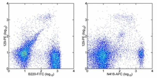
Applications Tested: This eBio129c (129c) antibody has been tested by flow cytometric analysis of SJL splenocytes. This can be used at less than or equal to 0.5 µg per test. A test is defined as the amount (µg) of antibody that will stain a cell sample in a final volume of 100 µL. Cell number should be determined empirically but can range from 10^5 to 10^8 cells/test. It is recommended that the antibody be carefully titrated for optimal performance in the assay of interest.
Excitation: 488-561 nm; Emission: 578 nm; Laser: Blue Laser, Green Laser, Yellow-Green Laser.
Filtration: 0.2 µm post-manufacturing filtered.
For Research Use Only. Not for use in diagnostic procedures. Not for resale without express authorization.
Product Information
Product Information
Shipping & Returns
Shipping & Returns
CD317 (BST2, PDCA-1) Monoclonal Antibody (eBio129c (129c)), PE, eBioscience
CD317 (BST2, PDCA-1) Monoclonal Antibody (eBio129c (129c)), PE, eBioscience
PRODUCT DETAILS
Host: Rat
Isotype: IgG2b, kappa
Clonality: Monoclonal
Clone: eBio129c (129c)
Format: PE
Reactivity: Ms
Application: Flow Cytometry
Tested Dilution: 0.5 µg/test
Concentration: 0.2 mg/mL
Storage: 4°C, store in dark, DO NOT FREEZE!
Formulation: PBS with 0.09% sodium azide; pH 7.2
Purification: Affinity chromatography
Data Sheet: TDS
Specific Information
Description: The eBio129c monoclonal antibody reacts with mouse PDCA-1 (BST2, CD317), a specific marker of plasmacytoid dendritic cells (pDC), also known as type I IFN-producing cells (IPC) in the naive mouse. Mouse IPCs are typically CD11c+, CD11b-, B220+, Ly-6C+, and CD62L+. PDCA-1 is predominantly expressed by IPCs in the naive mouse which represent sa very minor population (<0.5%) of splenocytes. It is upregulated on numerous cell types following stimulation which triggers an IFN response. PDCA-1 cycles between cell surface and intracellular compartments and may function to regulate trafficking of secreted cytokines. PDCA-1 (BST2) is the protein recognized by the antibody 120G8.
The epitope recognized by eBio129c is distinct from eBio927; thus, the antibodies can be used to costain, purify and identify pDCs.
Applications Reported: This eBio129c (129c) antibody has been reported for use in flow cytometric analysis.
Applications Tested: This eBio129c (129c) antibody has been tested by flow cytometric analysis of SJL splenocytes. This can be used at less than or equal to 0.5 µg per test. A test is defined as the amount (µg) of antibody that will stain a cell sample in a final volume of 100 µL. Cell number should be determined empirically but can range from 10^5 to 10^8 cells/test. It is recommended that the antibody be carefully titrated for optimal performance in the assay of interest.
Excitation: 488-561 nm; Emission: 578 nm; Laser: Blue Laser, Green Laser, Yellow-Green Laser.
Filtration: 0.2 µm post-manufacturing filtered.
For Research Use Only. Not for use in diagnostic procedures. Not for resale without express authorization.
Original: $432.00
-70%$432.00
$129.60Product Information
Product Information
Shipping & Returns
Shipping & Returns
Description
PRODUCT DETAILS
Host: Rat
Isotype: IgG2b, kappa
Clonality: Monoclonal
Clone: eBio129c (129c)
Format: PE
Reactivity: Ms
Application: Flow Cytometry
Tested Dilution: 0.5 µg/test
Concentration: 0.2 mg/mL
Storage: 4°C, store in dark, DO NOT FREEZE!
Formulation: PBS with 0.09% sodium azide; pH 7.2
Purification: Affinity chromatography
Data Sheet: TDS
Specific Information
Description: The eBio129c monoclonal antibody reacts with mouse PDCA-1 (BST2, CD317), a specific marker of plasmacytoid dendritic cells (pDC), also known as type I IFN-producing cells (IPC) in the naive mouse. Mouse IPCs are typically CD11c+, CD11b-, B220+, Ly-6C+, and CD62L+. PDCA-1 is predominantly expressed by IPCs in the naive mouse which represent sa very minor population (<0.5%) of splenocytes. It is upregulated on numerous cell types following stimulation which triggers an IFN response. PDCA-1 cycles between cell surface and intracellular compartments and may function to regulate trafficking of secreted cytokines. PDCA-1 (BST2) is the protein recognized by the antibody 120G8.
The epitope recognized by eBio129c is distinct from eBio927; thus, the antibodies can be used to costain, purify and identify pDCs.
Applications Reported: This eBio129c (129c) antibody has been reported for use in flow cytometric analysis.
Applications Tested: This eBio129c (129c) antibody has been tested by flow cytometric analysis of SJL splenocytes. This can be used at less than or equal to 0.5 µg per test. A test is defined as the amount (µg) of antibody that will stain a cell sample in a final volume of 100 µL. Cell number should be determined empirically but can range from 10^5 to 10^8 cells/test. It is recommended that the antibody be carefully titrated for optimal performance in the assay of interest.
Excitation: 488-561 nm; Emission: 578 nm; Laser: Blue Laser, Green Laser, Yellow-Green Laser.
Filtration: 0.2 µm post-manufacturing filtered.
For Research Use Only. Not for use in diagnostic procedures. Not for resale without express authorization.


