CD317 (BST2, PDCA-1) Monoclonal Antibody (26F8), PE, eBioscience
PRODUCT DETAILS
Host: Mouse
Isotype: IgG1, kappa
Clonality: Monoclonal
Clone: 26F8
Format: PE
Reactivity: Hu
Application: Flow Cytometry
Tested Dilution: 5 µL (0.25 µg)/test
Concentration: 5 μL/Test
Storage: 4°C, store in dark, DO NOT FREEZE!
Formulation: PBS with BSA and 0.09% sodium azide; pH 7.2
Purification: Affinity chromatography
Data Sheet: TDS
Specific Information
Description: This 26F8 monoclonal antibody reacts with human CD317 (also known as BST2 and tetherin). CD317 is a 30-36-kDa type II transmembrane protein expressed on B cells and bone marrow stromal cells. Although reports have indicated that CD317 mRNA is detectable in activated T cells, protein expression in freshly isolated primary T cells and macrophages is undetectable. Moreover, certain T cell lines, such as Jurkat, do not express detectable levels of CD317 protein. However, CD317 expression can be induced in many cell types (e.g., T and B cells, 293T, and HeLa) by IFNalpha treatment. CD317 has been associated with pre-B cell growth and the terminal differentiation of plasma B cells. More recently, this molecule has been reported to prevent HIV-1 virion release from the surface of infected cells, leading to reuptake and degradation of the virus. This activity is inhibited by the HIV-1 accessory protein Vpu. CD317 has been identified as the ligand for the ILT7 receptor.
Applications Reported: This 26F8 antibody has been reported for use in flow cytometric analysis.
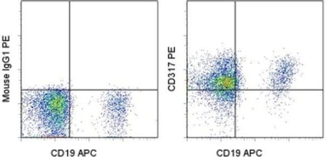
Applications Tested: This 26F8 antibody has been pre-titrated and tested by flow cytometric analysis of normal human peripheral blood cells. This can be used at 5 µL (0.25 µg) per test. A test is defined as the amount (µg) of antibody that will stain a cell sample in a final volume of 100 µL. Cell number should be determined empirically but can range from 10^5 to 10^8 cells/test.
Excitation: 488-561 nm; Emission: 578 nm; Laser: Blue Laser, Green Laser, Yellow-Green Laser.
Filtration: 0.2 µm post-manufacturing filtered.
For Research Use Only. Not for use in diagnostic procedures. Not for resale without express authorization.
Product Information
Product Information
Shipping & Returns
Shipping & Returns
CD317 (BST2, PDCA-1) Monoclonal Antibody (26F8), PE, eBioscience
CD317 (BST2, PDCA-1) Monoclonal Antibody (26F8), PE, eBioscience
PRODUCT DETAILS
Host: Mouse
Isotype: IgG1, kappa
Clonality: Monoclonal
Clone: 26F8
Format: PE
Reactivity: Hu
Application: Flow Cytometry
Tested Dilution: 5 µL (0.25 µg)/test
Concentration: 5 μL/Test
Storage: 4°C, store in dark, DO NOT FREEZE!
Formulation: PBS with BSA and 0.09% sodium azide; pH 7.2
Purification: Affinity chromatography
Data Sheet: TDS
Specific Information
Description: This 26F8 monoclonal antibody reacts with human CD317 (also known as BST2 and tetherin). CD317 is a 30-36-kDa type II transmembrane protein expressed on B cells and bone marrow stromal cells. Although reports have indicated that CD317 mRNA is detectable in activated T cells, protein expression in freshly isolated primary T cells and macrophages is undetectable. Moreover, certain T cell lines, such as Jurkat, do not express detectable levels of CD317 protein. However, CD317 expression can be induced in many cell types (e.g., T and B cells, 293T, and HeLa) by IFNalpha treatment. CD317 has been associated with pre-B cell growth and the terminal differentiation of plasma B cells. More recently, this molecule has been reported to prevent HIV-1 virion release from the surface of infected cells, leading to reuptake and degradation of the virus. This activity is inhibited by the HIV-1 accessory protein Vpu. CD317 has been identified as the ligand for the ILT7 receptor.
Applications Reported: This 26F8 antibody has been reported for use in flow cytometric analysis.
Applications Tested: This 26F8 antibody has been pre-titrated and tested by flow cytometric analysis of normal human peripheral blood cells. This can be used at 5 µL (0.25 µg) per test. A test is defined as the amount (µg) of antibody that will stain a cell sample in a final volume of 100 µL. Cell number should be determined empirically but can range from 10^5 to 10^8 cells/test.
Excitation: 488-561 nm; Emission: 578 nm; Laser: Blue Laser, Green Laser, Yellow-Green Laser.
Filtration: 0.2 µm post-manufacturing filtered.
For Research Use Only. Not for use in diagnostic procedures. Not for resale without express authorization.
Original: $375.00
-70%$375.00
$112.50Product Information
Product Information
Shipping & Returns
Shipping & Returns
Description
PRODUCT DETAILS
Host: Mouse
Isotype: IgG1, kappa
Clonality: Monoclonal
Clone: 26F8
Format: PE
Reactivity: Hu
Application: Flow Cytometry
Tested Dilution: 5 µL (0.25 µg)/test
Concentration: 5 μL/Test
Storage: 4°C, store in dark, DO NOT FREEZE!
Formulation: PBS with BSA and 0.09% sodium azide; pH 7.2
Purification: Affinity chromatography
Data Sheet: TDS
Specific Information
Description: This 26F8 monoclonal antibody reacts with human CD317 (also known as BST2 and tetherin). CD317 is a 30-36-kDa type II transmembrane protein expressed on B cells and bone marrow stromal cells. Although reports have indicated that CD317 mRNA is detectable in activated T cells, protein expression in freshly isolated primary T cells and macrophages is undetectable. Moreover, certain T cell lines, such as Jurkat, do not express detectable levels of CD317 protein. However, CD317 expression can be induced in many cell types (e.g., T and B cells, 293T, and HeLa) by IFNalpha treatment. CD317 has been associated with pre-B cell growth and the terminal differentiation of plasma B cells. More recently, this molecule has been reported to prevent HIV-1 virion release from the surface of infected cells, leading to reuptake and degradation of the virus. This activity is inhibited by the HIV-1 accessory protein Vpu. CD317 has been identified as the ligand for the ILT7 receptor.
Applications Reported: This 26F8 antibody has been reported for use in flow cytometric analysis.
Applications Tested: This 26F8 antibody has been pre-titrated and tested by flow cytometric analysis of normal human peripheral blood cells. This can be used at 5 µL (0.25 µg) per test. A test is defined as the amount (µg) of antibody that will stain a cell sample in a final volume of 100 µL. Cell number should be determined empirically but can range from 10^5 to 10^8 cells/test.
Excitation: 488-561 nm; Emission: 578 nm; Laser: Blue Laser, Green Laser, Yellow-Green Laser.
Filtration: 0.2 µm post-manufacturing filtered.
For Research Use Only. Not for use in diagnostic procedures. Not for resale without express authorization.


